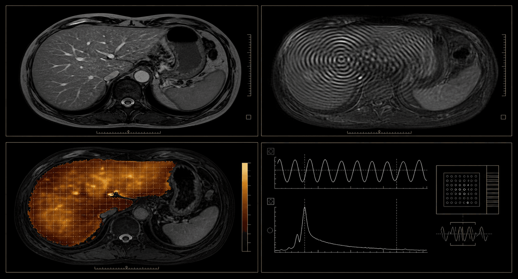
Why MRE must be matched to the examination
MRE is an MRI-based technique that estimates tissue stiffness. For a useful liver cohort, MRE wave images, stiffness maps or elastograms must be linked to the correct examination and matching anatomical sequences. A general abdominal MRI without verified MRE content is not an equivalent substitute.
Hospital inventory questions
The first hospital query asks for aggregate counts only. It checks disease labels, sequence combinations, scanner models, demographics and available clinical evidence before any export is requested.
- Number of patients and examinations for each priority liver disease
- Presence of MRE, T1-weighted, T2-weighted and DWI/ADC in the same study window
- Scanner manufacturer, model, field strength and protocol distribution
- Available reports, fibrosis stage, biopsy, laboratory results or other ground truth
- Age, sex and ethnicity fields where lawful and appropriately minimized
Feasibility before pricing
Release timing and commercial scope are established after inventory, missingness, privacy work and clinical-review burden are known. Programme records distinguish source imaging, derived MRE outputs, clinical truth and annotation work rather than compressing them into an unverified case count.
Questions, answered directly.
Is MRE the same as a normal liver MRI?+
MRE is performed on an MRI system but adds mechanical-wave acquisition and processing to estimate tissue stiffness. Not every liver MRI includes MRE.
What disease labels are most relevant?+
The programme prioritizes MASH/MASLD, viral hepatitis, cirrhosis, autoimmune hepatitis and primary sclerosing cholangitis.
How many cases are available?+
The programme is in sourcing. A verified count will be stated only after hospital inventory analysis.